A Glimpse Inside FVRC’s Internal Medicine Department
General

A Glimpse Inside FVRC’s Internal Medicine Department
Florida Veterinary Referral Center’s (FVRC) emergency and referral hospital comprises several different departments to help diagnose, treat, and manage difficult medical cases. Our internal medicine department is an integral hospital section that focuses on complex diseases that often affect multiple body systems, such as:
Gastrointestinal (GI) disease
Infectious disease
Respiratory disease
Cardiac disease
Urologic disease
Immune-mediated and hematologic disease
Endocrine disease
Musculoskeletal and neurologic disease
Cancer
Our board-certified veterinary internist, Dr. Amy Lang, collaborates with our surgery, radiology, emergency, and critical care departments to provide your pet with the best possible medical care. We also partner with other specialists and your family veterinarian to help diagnose and manage challenging cases that require specialized equipment, such as ultrasound, endoscopy, or advanced imaging.
Diagnostic Ultrasound for Pets
Ultrasound is a valuable diagnostic tool we use to observe your pet’s internal body structures in real-time to identify abnormal structures or malfunction. This versatile imaging modality allows us to evaluate your pet’s body areas, including:
Abdomen — Abdominal structures can be evaluated for proper anatomy and function. For example, we can use ultrasound to observe your pet’s intestines moving food through her GI tract, measure her stomach-wall thickness, or evaluate her gallbladder for stones.
Thorax — We can use ultrasound to visualize abnormalities in your pet’s thoracic cavity, such as a lung mass or enlarged lymph nodes.
Heart — An echocardiogram, which is an ultrasound examination of your pet’s heart function, can be used to measure heart-wall thickness and chamber size, observe blood flow, and evaluate valve function.
Urogenital structures — We can assess your pet’s urinary and reproductive systems for problems such as uterine cancer, prostatic enlargement, and kidney stones.
Eyes — We can perform an ocular ultrasound to examine your pet’s internal eye structures, and look behind her eyes for masses or swellings.
Ultrasound is also commonly used to obtain tissue samples during ultrasound-guided biopsies. Real-time imaging allows us to guide a needle directly to the site of concern and collect cells for microscopic examination to diagnose diseases.
Endoscopy in Pets
An endoscope is composed of a high-density camera on the end of rigid or flexible tubing that can be inserted into body cavities. The camera transmits an image to a monitor where we can see body structures in detail, allowing us to examine organs, collect tissue samples, or perform surgery. Endoscopy can be used in diagnostic testing, or as part of surgical treatment for some medical conditions.
As a diagnostic tool, endoscopy is used to examine internal body structures and collect tissue samples. An endoscope can be inserted into natural openings, such as the GI tract or the airways, or through a small incision into a joint or body cavity, such as the abdomen or thorax. Once inside the body, fluid or air can be used to expand the area for better visualization, and tools can be inserted through the endoscope tubing, or additional incisions, to grasp tissue samples.
Endoscopy is also used to perform minimally invasive surgery (MIS), which uses an endoscope and surgical tools inserted through several small incisions instead of opening up a body cavity with a single large incision. Since MIS uses smaller incisions, healing time and pain are significantly reduced, and your pet can return to normal function more quickly. MIS is commonly used to perform surgery of the abdomen, thorax, and joints. An endoscope can also be passed through a pet’s GI tract to retrieve ingested foreign objects, instead of making incisions through the abdominal cavity and GI structures.
Advanced Imaging for Pets
We offer a number of advanced imaging modalities to obtain diagnostic images, including:
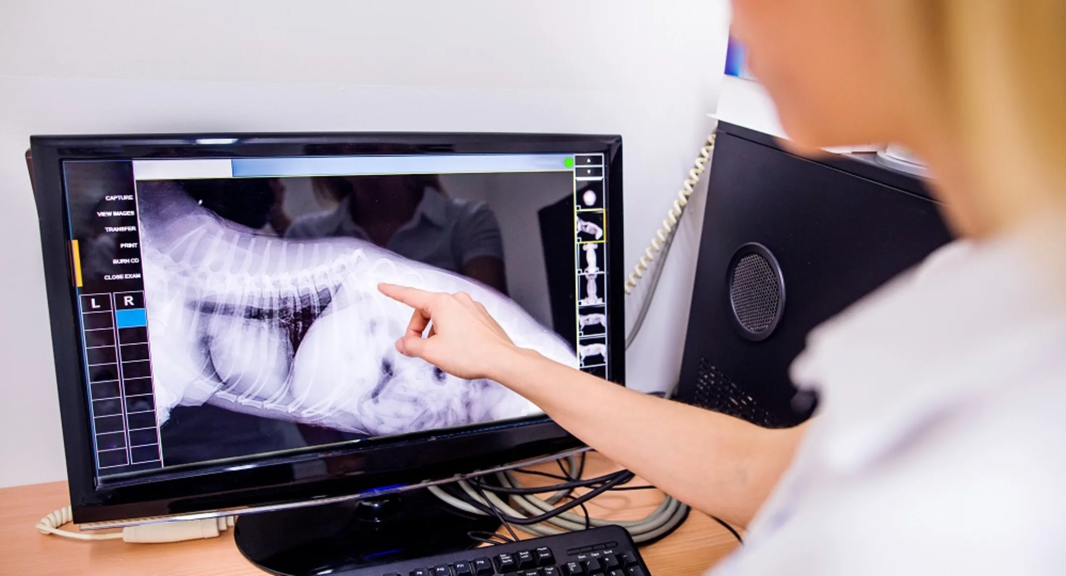
Digital radiography — A digital radiography unit takes X-rays that are transmitted to a computer for viewing, rather than a printed piece of film. We can easily send your pet’s digital X-rays to your family veterinarian to keep her informed about your pet’s diagnosis.
Computed tomography (CT) — A CT scan gathers images of a body area in small slices, which are reconstructed to obtain a three-dimensional image. CT scans provide greater detail than digital X-rays, and superior images of soft-tissue and bony structures. CT has a variety of uses, such as locating tumors to plan a surgical approach, identifying the cause of chronic nasal discharge, and screening for cancer metastasis.
Magnetic resonance imaging (MRI) — MRI is the most advanced imaging technique available in human and veterinary medicine. MRI provides the most detailed images of nervous system structures, including the brain and spinal cord, and is considered the gold standard for imaging these structures.
If you have questions about our internal medicine department’s services, or if your family veterinarian has referred you to FVRC for advanced diagnostic or treatment options, contact us.
